
 1900 1717
 The heart can be said to be an extremely important organ in the human body. The main function is in charge of providing oxygen and nutrients for the working organs in the body, and at the same time removing waste products during metabolism. And to detect abnormalities in this organ, the echocardiography method is remarkable scientific progress to check and diagnose heart diseases.
Since then, giving timely treatment to ensure the human heart is always healthy. So what is echocardiography? Let's learn the basics of echocardiography right now.
1. What Is Echocardiography?
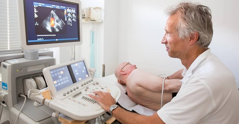
Echocardiography is a method of probing and diagnosing using high-frequency ultrasound waves to show real pictures of the heart and its related structures. This method is a non-radiative, non-invasive technique with rare side effects.
During an echocardiogram, the doctor can examine and evaluate problems such as heart wall size and thickness, heart valve activity, damaged or weakened areas of the heart muscle. In addition, echocardiography is also used in periodic health checks, re-evaluating the patient's condition after a heart attack or stroke.
2. What Are The Types of Echocardiography?
Currently, there are the following types of echocardiography:
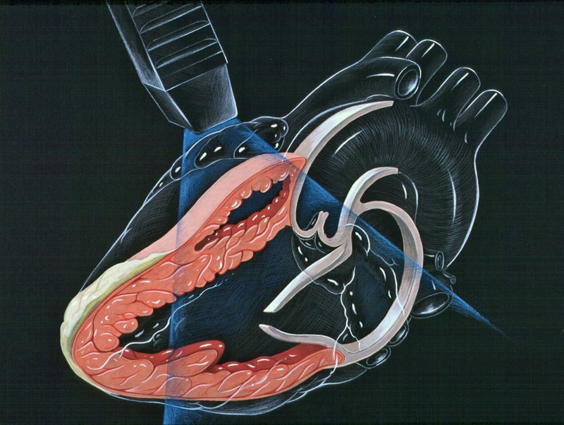
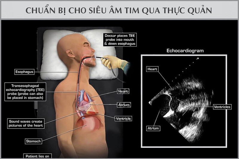
Transesophageal echocardiography: Using a thin end attached directly to the end of the endoscope and into the esophagus to better observe the details of the heart organ from the back. This method provides more detailed images than the traditional method.
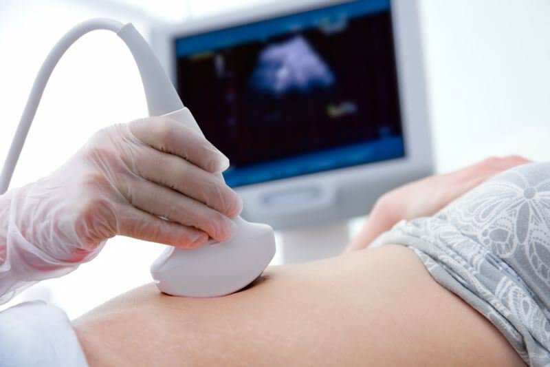
Chest wall echocardiography: This is the most common method, which is performed by using a solid probe on the chest part of the body near the heart. There is a layer of gel applied to the skin for better sound wave transmission.
- Doppler ultrasound: This ultrasound helps check the amount of machine and pressure from the pulmonary artery. Your doctor can also use this method to map the speed and direction of blood flow in your heart. The results of Doppler ultrasound can detect a number of diseases of the heart and blood vessels.
- Echocardiogram: Can walk or run on the treadmill. Your doctor will monitor your heart rate and blood pressure during pre-and post-exercise checks. This method can help diagnose conditions such as myocardial ischemia, heart failure, and heart valve problems.
- Three-dimensional echocardiography: Also known as 3D ultrasound, creating detailed images of the heart's chamber. Imaging helps assess heart valve function in people with heart failure. Diagnosis of congenital heart problems in a newborn. Evaluate heart function, check structure before heart valve replacement surgery, or some other intervention surgeries.
- Ultrasound of the fetal heart: The doctor can use the method of ultrasound of the fetus to check the activity of the fetal heart. Usually, this method is done when the baby is about 18 to 22 weeks old.
3. What Can Echocardiography Detect?
In the past, echocardiography was not widely used because the machines were bulky, difficult to perform, and expensive. However, with the development of science and technology today, this method has become popular and is one of the factors that help doctors diagnose heart diseases more accurately.
The doctor can observe the heart's structure, the way the heart contracts, the heart valves work, the shape of the heart. Through it, the doctor can detect and diagnose pathological problems in the heart, including:
- Stenosis of the heart valve, open-heart valve: The disease occurs when the structure of the heart valve is deformed, the valve is not closed, leading to blood circulation to the heart.
Change in heart size: abnormally sized chambers and heart muscles due to high blood pressure or some other medical condition.
Damaged heart muscle: abnormalities in the ejection process are detected to help prevent dangerous complications such as acute myocardial infarction.
Heart defects: This method helps determine if the heart has birth defects or not in infants and young children.
Pericardial effusion: Pericardial effusion, if not detected and treated in time, can lead to heart failure.
- Monitoring the treatment methods of heart diseases: Monitoring the response of the heart organ to different heart treatments.
4. Which Case Need An Echocardiogram?
- There are many reasons for a person to perform an echocardiogram. Maybe when detecting signs of abnormal cardiovascular through other tests. Or perhaps through a stethoscope and heart disease manifestations.
- In case the patient has a suspected problem related to the heart organ with the following symptoms, it is best to have an echocardiogram for timely diagnosis:
- Chest pain with dizziness and headache.
- Fast and slow heart rate, standard arrhythmia, difficulty breathing.
- Suspected heart disease due to a relative having a history of cardiovascular disease.
- Heart palpitations, difficulty breathing, or shortness of breath when doing hard work.
- Chest pain, shortness of breath, unusual shortness of breath while living.
- Some manifestations of myocardial ischemia such as neck pain, arm pain, back pain, jaw pain also need echocardiography.

5. What Problems Should Be Noted During Echocardiography?
This is a modern method of examination, so the patient does not need any special preparation before performing an ultrasound. For normal echocardiography, the patient can eat and drink normally before the procedure. If an EEE or stress ultrasound, the patient may be told not to eat or drink for several hours before the scan.
The patient's throat will be numbed with a spray tube to make it easier to pass the transducer through the esophagus. So you will need a sedative to help you relax. In case the doctor will ask the patient to stay in the hospital or clinic to monitor the health after completing the examination.
The transesophageal echocardiogram is due to the main sedative, so you won't be able to drive the motorbike yourself afterward. It is best to travel with loved ones or take public transport.
Before an echocardiogram, the patient will be asked to undress, the doctor will use a detector with ultrasound waves moving toward the heart. The patient will lie on the left side, the doctor will apply a gel layer on the area where the ultrasound probe is exposed to limit the air between the head and the patient's skin.
The ultrasound results will appear on the screen in black and white or in color. Time to perform in about 15-30 minutes as directed by your doctor.
6. What Is The Difference Between Echocardiography And Ultrasound?
Each echocardiogram has different functions. Specifically:
Stress ultrasound: When measuring an electrocardiogram or giving the patient medications that can make the heart beat faster, stronger.
Doppler ultrasound: Used to measure blood flow velocity at different locations in the heart chamber.
Transesophageal Ultrasound: The patient will swallow one end of a probe with a thin optical cable connected to the ultrasound.
7. Some Side Effects And Complications of Echocardiography
Echocardiography is a method of diagnosing diseases of the heart and related modern organs for accurate results. Above all, this method is painless, with no complications left. However, there are some rare but possible side effects such as:
- In cases where electrodes are attached to monitor the electrocardiogram at the same time, the patient will feel a bit uncomfortable when removing the tape for electrodes on the body.
A transesophageal echocardiogram can make your throat feel uncomfortable for a few hours. The possibility of internal throat scratching is absent or very rare. During the ultrasound, the patient may experience some breathing problems caused by sedatives.
- In the case of drug or exercise in cardiac ultrasound exercise can cause temporary arrhythmia. However, this is not caused by an echocardiogram.
After an echocardiogram, the patient can resume their usual work. In the event that an echocardiogram is normal, with a healthy heart, no further testing is needed. If in the case of negative results, the patient will continue to check and treat as required by the doctor.
Current echocardiography is a fairly safe and popular method. And there is no scientific basis to indicate a disadvantage in echocardiography. Therefore, if you feel any cardiac abnormalities or echocardiography immediately. Modern machinery and equipment will help doctors quickly diagnose, accurately, and promptly give treatment measures.
Above is the content of echocardiography, hope to bring you the most useful information to help equip you and your family with knowledge about echocardiography. As well as diseases related to heart organs need timely diagnosis and treatment. If you have any unusual symptoms, you need to go to a reputable hospital for an ultrasound, test for early treatment.
For more information about our test menu and price list, please click here.
The site cannot and does not contain medical advice. The medical information is provided for general informational and educational purposes only and is not a substitute for professional advice. Accordingly, before taking any actions based upon such information, we encourage you to consult with the appropriate professionals.